ADDED Tier-2 · Quick Summary
Quick Summary
Most patients recover from MPFL reconstruction in 6 to 12 months, with daily-activity comfort returning around weeks 6 to 12 and full sport-specific clearance typically between months 4 and 6. High-impact athletes may need 12 to 18 months. Recovery advances by milestones — quadriceps strength, range of motion, and stability — not by the calendar alone. Strict adherence to physical therapy is the single biggest predictor of a successful outcome.
Wondering when you’ll fully recover from medial patellofemoral ligament (MPFL) reconstruction? You’re not alone. Whether you’re an athlete, a parent of a young patient, or anyone eager to return to a pain-free, active lifestyle, understanding how long the MPFL recovery process will take is important. This guide draws on my expertise and provides direct links to detailed resources, so you can equip yourself with the information that will help you recover with minimal confusion or stress.
What is MPFL Reconstruction?
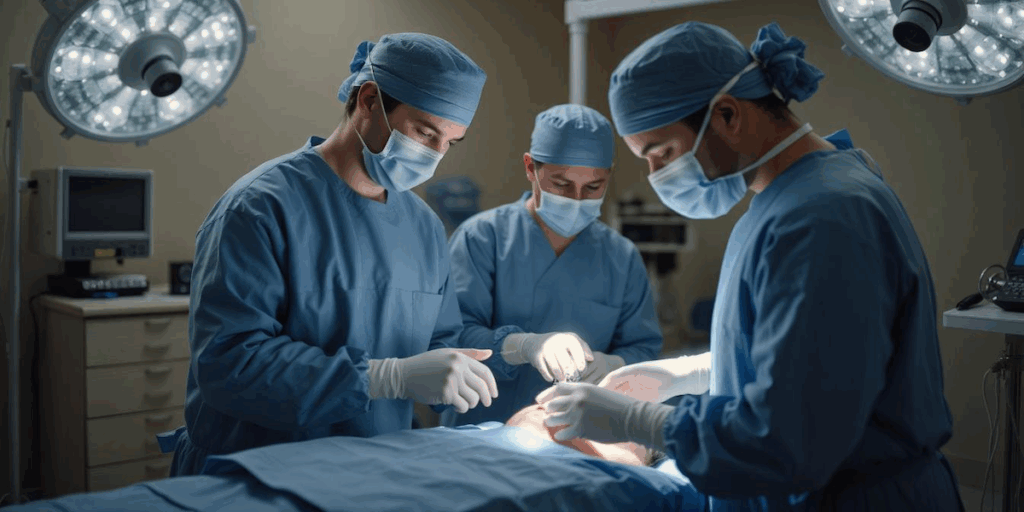
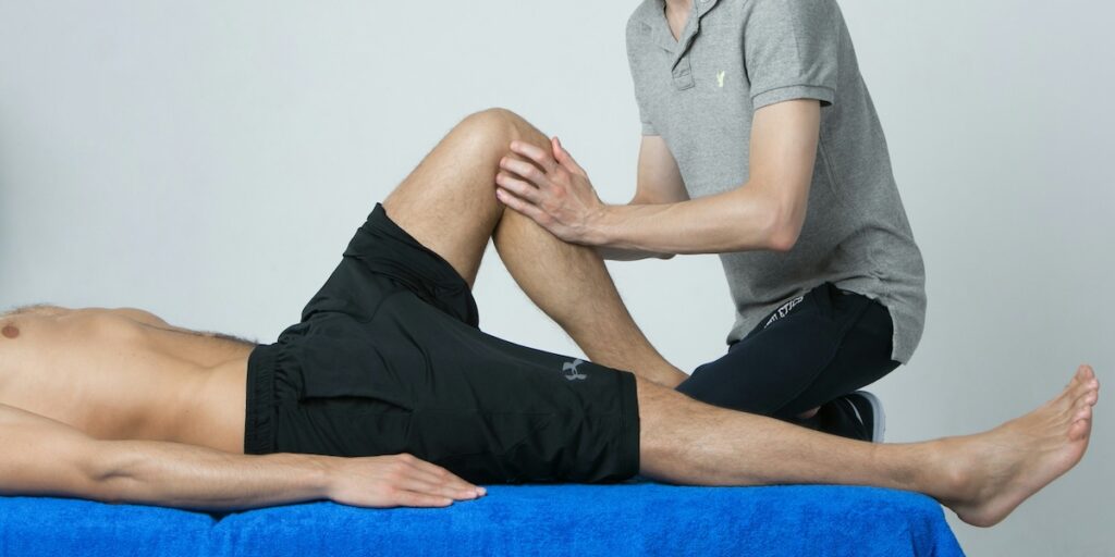
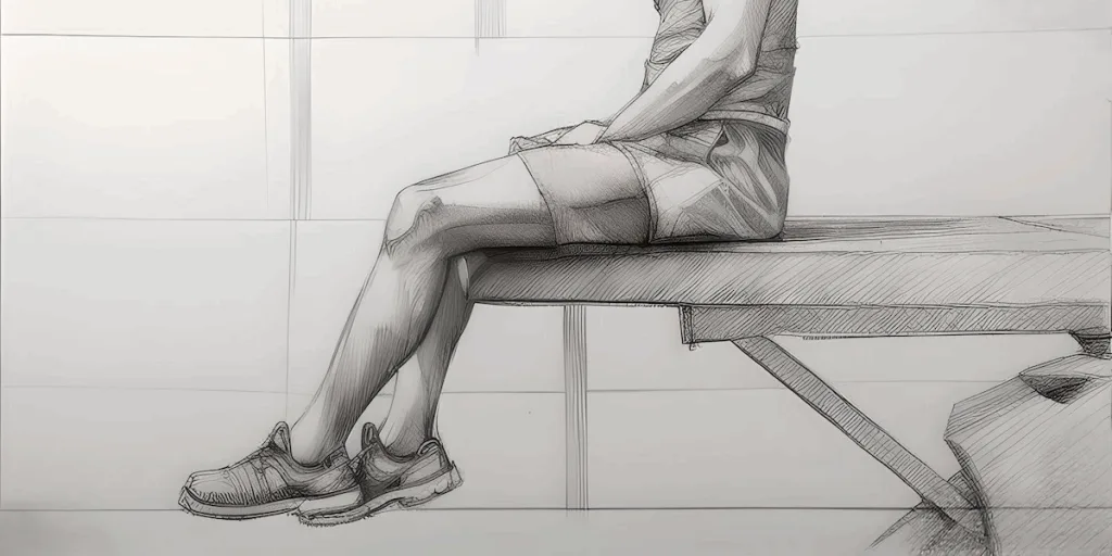
MPFL reconstruction is a surgical procedure designed to restore stability to the kneecap (patella) after a patellar dislocation or chronic instability. The surgery rebuilds the medial patellofemoral ligament — the soft-tissue tether on the inner side of the knee that keeps the patella in its trochlear groove — using a graft (commonly from a hamstring tendon) anchored between the femur and patella. This ligament is essential for normal knee function and preventing future dislocations.
If you have additional procedures performed alongside MPFL reconstruction (such as a tibial tubercle osteotomy), your MPFL recovery timeline and rehabilitation recommendations may differ somewhat. Be sure to discuss your individualized protocol with your surgical team.
Remember, advancing from one phase to the next is based not only on time but also on meeting specific clinical and functional milestones. Your surgeon or therapist will assess your strength, range of motion, and pain levels before clearing you to move forward.
MPFL Recovery Timeline
The timeframes that follow are general guidelines. Your specific plan may change, especially if you have additional procedures. Always follow your care team’s personalized instructions.
Phase 1: Immediate Post-Operative (Weeks 1–2)
Goal: Minimize swelling, protect surgical repair, begin gentle movement.
What to expect:
- Use of crutches and a knee brace
- Protected weight-bearing on the operated leg
- Gentle range-of-motion exercises
- Return to sedentary work or school is possible within a week depending on how much walking is required
Practical tips for the first few weeks:
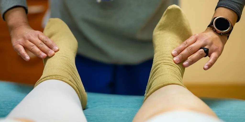
- The leg is placed in a brace for up to six weeks to keep it straight during walking until the quad is strong enough to control the leg; follow all instructions for use. For some patients, the brace is worn nearly full-time (except for physical therapy or hygiene reasons), per your surgeon’s orders.
- Begin rehabilitation as directed to restore quadriceps strength, which is essential for stability and safe brace removal.
- Patients can walk with weight on the operated leg immediately, while wearing the brace.
- Devices, including muscle stimulators (for example, Zynex NexWave | Prescription Pain Management Tens Unit) and cold compression machines (such as the NICE, available in my recovery shop) can help reduce swelling and pain and support muscle recovery.
- Strictly follow all post-operative instructions, especially any restrictions on knee movement or activity
Phase 2: Early Rehabilitation (Weeks 3–6)
Goal: Increase range of motion, start muscle activation, decrease use of crutches.
What to expect: Under the guidance of a physical therapist, strength and motion work intensifies. You’ll gradually transition to out of the brace as tolerated.
Practical tips for weeks 3-6:
- The brace should remain on during walking and other weight-bearing activities until you have good quad control (ask your physical therapist).
- Begin bending and straightening the knee as recommended by the physical therapist, but only within the limits set by the surgeon to protect healing tissues.
- Patients usually bear full weight on the leg while using the brace. Be cautious and avoid activities that may risk a slip or fall.
- Engage in prescribed exercises to activate and strengthen the quadriceps, which helps prepare for brace removal and future mobility.
- Continue to use cold therapy devices and muscle stimulation devices (if recommended) to reduce inflammation and support healing.
- Attend all physical therapy sessions, perform home exercises, and promptly communicate any new or worsening symptoms, such as swelling, pain, or instability, to your care team. Staying consistent and proactive is key to a safe recovery.
- Refrain from running, jumping, or rapid direction changes during this phase to protect the new ligament and healing tissues.
Phase 3: Advanced Strengthening (Weeks 7–12)
Goal: Recover muscle strength, balance, and coordination.
What to expect: Start low-impact activities such as stationary cycling, elliptical, and bodyweight exercises. You can also slowly return to household chores and basic activities.
Practical tips for weeks 7-12:
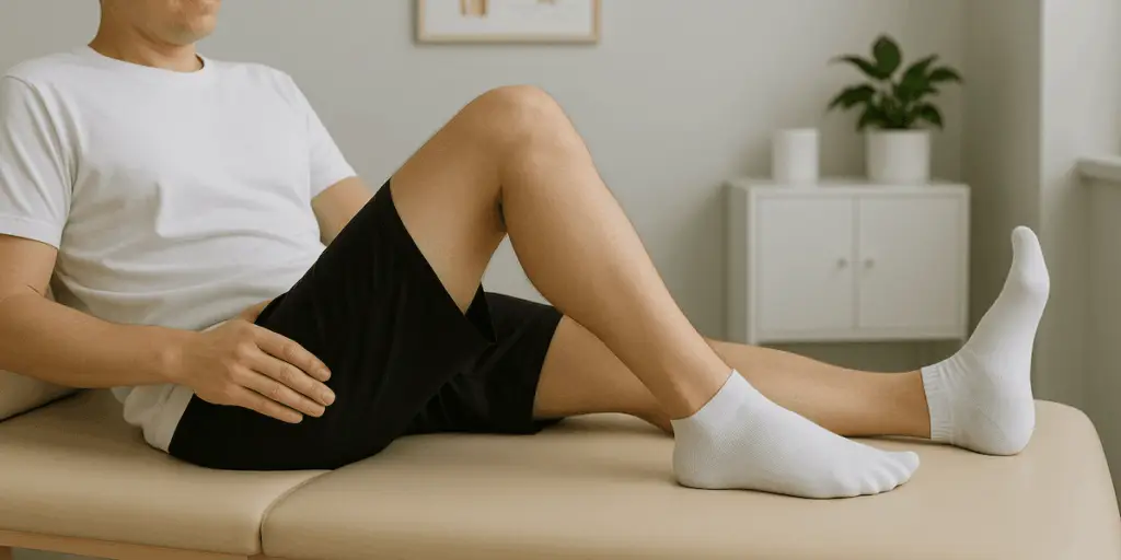
- As approved by your surgeon, discontinue the knee brace as your quadriceps strength and stability improve.
- Work with the physical therapist to restore full knee bending and straightening, advancing the range of motion exercises as tolerated and cleared.
- Transition to more challenging strengthening exercises for the quadriceps, hamstrings, and hip muscles, incorporating light resistance and functional movements.
- Begin balance drills and proprioceptive activities (such as standing on one leg or both legs on a bosu ball) to enhance joint stability and neuromuscular control.
- Activities such as stationary cycling, elliptical, and swimming (if cleared) can improve fitness without risking the healing ligament.
- Continue to watch for swelling, pain, or instability. Report any concerning changes to your orthopedic team promptly.
- Continue to avoid high-impact activity, such as running, jumping, and pivoting even if the knee feels stronger, to ensure safe healing of the repaired ligament.
- Maintain regular physical therapy visits and perform prescribed home exercises daily to support your ongoing recovery progress.
- Understand that healing is continuing; pushing too hard or rushing this phase can jeopardize your surgical outcome.
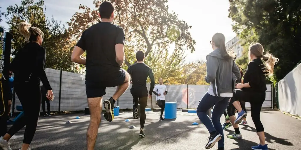
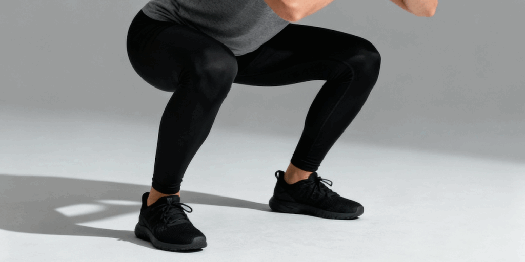
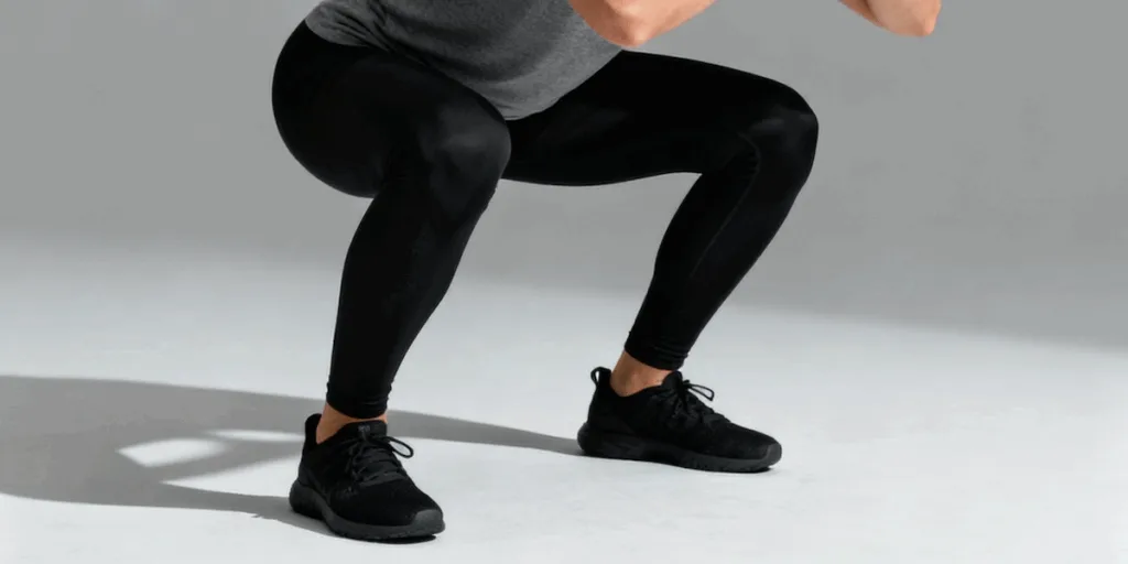
Phase 4: Functional and Sport-Specific Training (Months 3–4)
Goal: Prepare the knee for higher loads and dynamic movement.
What to expect: At this point, you’ll probably be able to add jogging on flat surfaces, agility drills, and proprioception training. For athletes, now is the time to lay the foundation for a gradual return to practice.
Practical tips for months 3-4:
- Under the guidance of the physical therapist, begin light plyometric (jumping) exercises, agility drills, and sport-specific movements as your strength, balance, and confidence improve.
- Initiate a graduated running program, starting with straight-line jogging before moving on to cutting, pivoting, or directional changes, which you should start only when cleared by the surgeon or physical therapist.
- Maintain a comprehensive strengthening program for the quadriceps, hamstrings, hips, and core to ensure muscular balance and protect the knee during dynamic activities.
- Incorporate advanced balance, coordination, and single-leg activities to prepare for unpredictable environments.
- Stay alert for pain, swelling, or instability during higher-intensity training. If it occurs, stop the activity and notify your orthopedic surgeon or physical therapist for assessment and guidance. These symptoms can indicate overuse, or a potential complication such as ligament failure or graft irritation.
- If you’re an athlete, progressively add drills that mimic the movements and demands of your particular sport, focusing first on technique before increasing speed and intensity.
- Participate in functional and readiness assessments as directed by the rehabilitation team to ensure it’s safe to return to competitive sports.
- Don’t advance to full participation until cleared; early return increases the risk of reinjury or graft failure.
- Keep open communication with your surgeon, physical therapist, and coach to adjust training and address concerns throughout your transition back to sport.
Phase 5: Return to Full Activity (Months 4–6+)
Goal: Restore full function, resume normal sports and life activities.
What to expect: This is the phase you’ve been working towards. You can now return to running, jumping, and contact sports as cleared by your surgeon and physical therapist. Typically, safe return to sport is considered only when your surgical leg matches or exceeds 90% of your non-operative leg in strength and hop tests, in addition to demonstrating good balance and confidence during sport-specific movements. It’s normal for recovery to include occasional setbacks, such as episodes of stiffness, discomfort, or slower progress. Communicate any concerns to your care team promptly. Most issues can be managed with adjustments to your rehabilitation plan.
Practical tips for months 4-6+:
- Ensure that you pass all return-to-play functional and strength tests before fully resuming sports participation, as advised by your surgeon or physical therapist.
- Maintain a consistent training program for leg strength, endurance, balance, and core stability, while closely monitoring for any renewed pain, swelling, or instability. If symptoms return, reduce activity and consult your care team before resuming higher-intensity movements.
- Gradually increase training intensity, volume, and sport-specific drills. Don’t jump back to full competition immediately.
- Incorporate a structured warm-up before every session and prioritize recovery strategies, such as stretching and ice, after high-intensity activity.
- Consider using supportive gear (such as knee sleeves or braces) if recommended during initial return to high-risk sports or movements.
- Remember, recovery is unique for each person. Don’t rush your process. Safe progression depends on milestone achievement, not just the passage of time or comparison to others. Listen closely to your body and your care team.
When You Are “Fully Recovered” from MPFL Reconstruction
Once you’ve met these important recovery benchmarks, continued maintenance and self-monitoring are essential for keeping your knee strong and healthy. Here’s what to expect:
- Pain, swelling, or instability should be fully resolved.
- Range of motion and strength should closely match your non-operative side.
- You are able to perform all work, sports, or daily activities with confidence. Often, this happens between 6 to 12 months after surgery. However, high-impact athletes may need up to 12–18 months for full sports readiness.
Even after achieving these benchmarks, it’s important to continue working on strength, flexibility, and endurance to protect your knee and support long-term recovery. Maintaining these habits will help you prevent reinjury and support your return to all activities.
Patient Stories and Success
Hear from patients who’ve completely recovered from MPFL reconstruction surgery—what worked for them, lessons they learned, and words of encouragement:
Pro Tips for Optimal MPFL Recovery
- Stick to your rehab plan: Commitment to a structured rehabilitation program is the top predictor of success.
- Be patient and proactive: Some days will be easier than others; don’t get discouraged. Tracking your progress in a document can help you see the milestones you’ve already achieved.
- Communicate: Keep your surgeon and physical therapist informed about any new pain, swelling, or concerns.
- Prioritize mental readiness: Addressing confidence and emotional well-being is often just as important as physical recovery. Talk to your care team if you have any anxiety or concerns about returning to sport or activity or seek out help from a sports psychologist as they can really help your on your road to recovery.
These guidelines provide a general road map, but always rely on recommendations from your surgical team and physical therapist for guidance tailored specifically to you.
Accessing Expert Help and Next Steps
Recovering from MPFL reconstruction is a gradual journey that requires dedication, patience, and close partnership with a skilled care team. By following a structured timeline, embracing rehabilitation, and listening to your body through each phase, you can maximize your chances of regaining strength, stability, and confidence. With thoughtful guidance and the right resources, most people can safely return to their favorite sports and activities, enjoying renewed mobility for years to come.
If you have questions about whether an MPFL reconstruction is right for you or want a second opinion, please reach out.
Frequently Asked Questions About MPFL Recovery
How long does it take to recover from MPFL reconstruction surgery?
Most patients return to daily activities within 6 to 12 weeks after MPFL reconstruction, with full sport-specific recovery typically taking 6 to 12 months. High-impact athletes may need 12 to 18 months for full sports readiness. Recovery is based on hitting milestones, not just on the calendar — your surgical team will check strength, range of motion, and stability before clearing you to move through each phase.
How soon can I walk after MPFL reconstruction?
Patients can usually put weight on the operated leg right after MPFL reconstruction, while wearing a knee brace. The brace keeps the leg straight during walking until the quad muscle is strong enough to control the knee — usually for up to six weeks. Crutches are used for extra support and balance during the first few weeks.
When can I return to sports after MPFL reconstruction?
Return to sport is usually considered between 4 and 6 months after MPFL reconstruction, but only when your surgical leg matches or exceeds 90% of the strength and hop-test performance of your other leg. High-impact and contact athletes may need up to 12 to 18 months. Functional readiness — not the calendar — determines a safe return to play.
How long do I need to wear a knee brace after MPFL surgery?
Most patients wear the knee brace for up to six weeks after MPFL reconstruction, mainly during walking and weight-bearing activity. The brace keeps the knee straight until your quad strength is enough to stabilize the joint on its own. Your surgeon will clear you to stop using the brace once you show good quad control.
Is MPFL reconstruction painful during recovery?
Most patients report manageable post-surgery discomfort that improves significantly within the first 1 to 2 weeks. Pain is controlled with prescribed medication, cold-compression devices, and muscle stimulators. Sharp pain, increasing swelling, or new instability beyond the first weeks should be reported to your surgical team right away — these can signal a complication.
What slows down MPFL recovery the most?
The two biggest things that slow MPFL recovery are skipping physical therapy sessions and rushing back to high-impact activity before clearance. Missing rehab sessions delays the quad strength gains needed to come out of the brace and progress. Going back to running, jumping, or pivoting before milestone clearance increases the risk of the graft failing or the kneecap dislocating again.